Imaging technology is critical to the field of dentistry, but it has seen many recent advances because of technological improvement. The quality, convenience safety, and accuracy have all been upgraded. Visits to the dentist are now routine with x-rays conducted in just a few seconds.
These advancements took place largely within the last hundred years which is not surprising because there have been similar advancements within the entire field of modern medicine. There is a lot of research highlighting the countless benefits of medical imaging technology.
It is an example of technology that is integral to healthcare. These upgrades can be attributed to modern tools and equipment that dental practitioners are able to use which give them access to parts of the anatomy that were out of their reach. The ability to use upgraded imaging is an invaluable tool when it comes to dentistry.
Dentists would be unable to detect serious diseases and the exact location of impaired teeth if it were not for the advances in imaging technology. Mankind has indeed come a long way from equipment that would have dangerous consequences, like irreparable damage to the skin and hair. This would be because of dangerous radiation levels that came along with x-rays. protective equipment would be used for safety, but the damage would still occur.
In consultation with experienced dentist Dr. Ronkin, we uncovered a few of the many improvements to imaging as well as new approaches and that are practiced because of this upgraded technology. For example, sentences are now able to access images of inside and outside the mouth before a course of action is taken to treat problems. This provides greater levels of comfort to the patients and makes the dentist’s job easier.
In the last three decades, the advancement in imaging technology has been invaluable to dental practitioners globally. So much so that the way dentistry is taught also needed to be changed, so then these advancements can be accommodated. Students now need to learn how to use the most accurate diagnostic tools and special methods of imaging which are commonplace in the dental field.
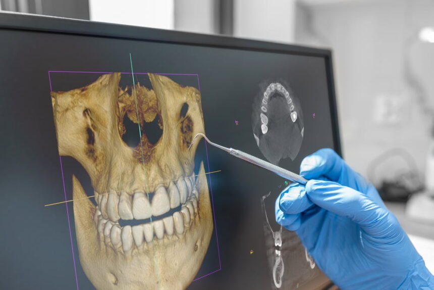
These imaging methods include ultrasound, magnetic resonance imaging, computed tomography, and much more. Because of the use of these technologies, dental practitioners can now store the images of all of their patients as well as color and control the brightness and contrast. The images are able to be displayed in 3D format giving a clearer view of problem areas.
This greatly helps the dentist who can now detect issues like decay and cavities as well as the progression of children’s teeth. They can make critical decisions as it pertains to treatment and give an early diagnosis when they encounter any abnormalities. Patients are able to benefit from a more competent dentist who can spot tumors, cysts, and abscesses in the mouth. Dentists are also able to solve many problems because of the use of dental imaging such as dental implants, root canal treatment, oral surgery, and much more.
Ultrasound – This Imaging method is inexpensive and non-invasive. Ultrasounds are also painless without using ionizing radiation which can be quite harmful. You can use ultrasounds for both soft and hard tissue in the mouth. Ultrasounds use the reflection of sound waves with frequencies that are outside the range that human beings can hear. The echoes are collected and converted into electrical signals to be displayed on a computer screen.
With ultrasounds, dental practitioners can detect extracapsular tractors as well as maxillofacial fractures. It is used in the diagnosis of fractures in the orbital margin anterior wall of the frontal sinus and nasal bone.
Magnetic Resonance Imaging or MRI – MRI’s are perhaps the most effective and widely used imaging technology for all viewing soft tissues in the anatomy without the need for invasive procedures. It also has the benefit of not using ionizing radiation. Most MRI machines are graded based on their magnetic strength which is measured in Tesla units. MRIs involve the use of hydrogen atoms within a magnetic field which create a magnetic resonance image.
One of the major benefits of MRIs is that a direct multiplanar image can be achieved without reorienting the patient. You can also grab the best resolution with low inherent contrast when it comes to human tissue. Dentists use it to look at soft tissue lesions in the salivary glands.
Computed Tomography or CT – A CT scanner uses a radiographic tube that is attached to ionization chambers or scintillation detectors. Modern CT scanners are able to reduce the amount of space and time and also reduce artifacts. They can give submillimeter resolution and up to 0.4 mm. These are very costly machines, however, so it would be a major investment for a dental practice.
CT scanners were the first imaging technology that allowed dentists to see soft and hard tissues of the facial bones when scanning the mouth. With image processing enhancement they could see cross-sectional images that were non-superimposed. Some CT programs are even able to highlight lesions using color enhancement.
To Sum It Up
The field of dentistry has improved considerably because of the advances made in imaging technology. Dental treatment and diagnostics have been revolutionized so that dental visits are faster and more cost-effective. It is also less harmful because of improved radiographic techniques. These changes have helped to reduce mortality while improving the quality of life for everyone.


