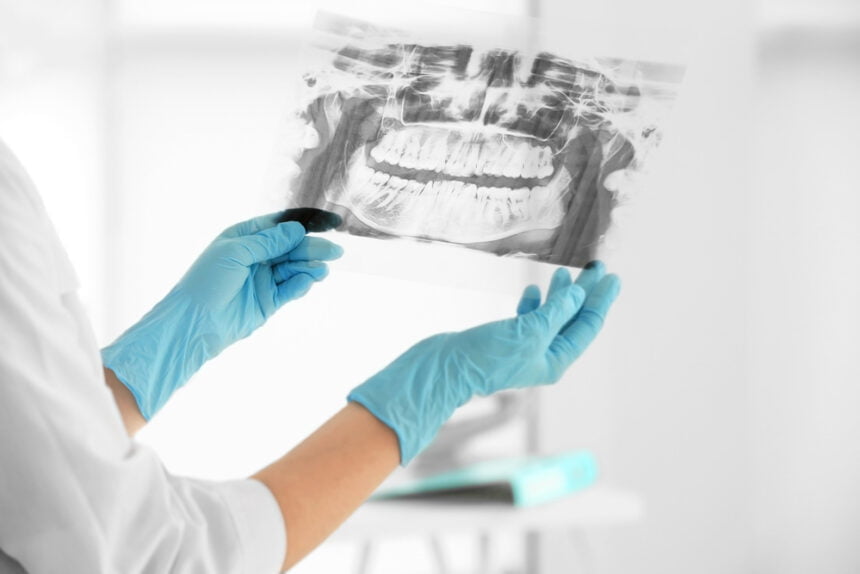
Dentists utilize dental X-rays, commonly referred to as dental radiography, to create pictures of the teeth, bones, and soft tissues around the mouth.
Dental practitioners may uncover problems that might not be obvious to the human eye by using X-rays, which emit a little quantity of radiation to create pictures.
We learned from Green Apple Dental that in order to prevent, diagnose, and treat dental diseases, dental X-rays are a crucial tool.
They might uncover unnoticed oral issues such as bone loss, tooth decay, cavities, and gum disease.
Dental X-rays may aid in preventing the emergence of more significant and expensive dental diseases by spotting concerns early. Depending on your particular dental requirements, your dentist may choose to employ a variety of dental X-rays.
Intraoral X-Rays
Dental X-rays are often obtained intraorally, or inside the mouth. In this way, dental problems that may have been missed during a visual examination may be identified and treated. Various intraoral X-rays exist, like:
- Bitewing X-rays: These X-rays reveal the crowns (the portion of the tooth that protrudes above the gumline) of the upper and lower teeth and may reveal cavities between teeth.
- Periapical X-rays are used to identify issues including abscesses, impacted teeth, and bone loss since they reveal the whole tooth, from the crown to the root.
- Occlusal X-rays: These X-rays are used to identify developmental issues and find foreign objects, and they reveal the complete arch of teeth in either the upper or lower jaw.
Diagnosing and treating dental problems including cavities, gum disease, and root canal infections requires the use of intraoral X-rays. They help find oral cancer and track how a child’s teeth develop. However, they are seldom used since they expose patients to more radiation than traditional dental X-rays.
Extraoral X-Rays
By contrast, extraoral X-rays are taken away from the mouth and may reveal more of the head and neck. Jaw, temporomandibular joint (TMJ), and facial bone problems may all be identified with their help. Several different kinds of X-rays may be taken outside the mouth.
- In a panoramic X-ray, the upper and lower jaws, teeth, and supporting structures are all seen in one image. Impacted teeth, tumors, cysts, and jawbone issues may all be seen with the use of one of these instruments.
- Cephalometric X-rays: These X-rays are used to assess the location, growth, and development of the jaw, and they offer a side image of the face. Overbites, underbites, and crossbites are just some of the orthodontic problems that may be detected with their help.
- Tomograms are cross-sectional X-rays of the jawbone that help doctors discover conditions including fractures, tumors, and infections.
Although not as detailed as intraoral X-rays, extraoral X-rays are nonetheless helpful for identifying issues in the head and neck because of the greater area they cover. When repeated X-rays are required, they are a safer alternative to intraoral X-rays since they expose patients to less radiation.
Panoramic X-Rays
If you need an X-ray of your mouth, namely the upper and lower jaws, teeth, and surrounding tissues, you may want to consider getting a panoramic X-ray. Dentists often utilize them to identify and treat a wide variety of dental issues, such as:
- Panoramic X-rays may reveal impacted teeth, or those that are not erupting normally from the gums.
- These X-rays are useful for diagnosing jaw issues such cysts, tumors, and bone loss.
- Planning orthodontic treatment with braces, Invisalign, or other orthodontic products requires panoramic X-rays.
- Pain and difficulty chewing are symptoms of temporomandibular joint (TMJ) abnormalities, which may be seen on panoramic X-rays.
- Oral cancer: These X-rays may spot tumors and lesions of oral cancer in their earliest, most curable stages.
Panoramic X-rays are faster and more convenient than traditional X-rays since they don’t need any preparation on the patient’s part and show the whole mouth in a single picture. They are a useful diagnostic tool for dentists due to the minimal amount of radiation they emit.
Cone Beam Computed Tomography (CBCT)
Dental CBCT, or cone beam computed tomography, provides high-resolution 3D pictures of the jaw, face, and teeth. Multiple views of the patient’s head are taken using a cone-shaped X-ray beam that revolves around the body. After combining these photos, dentists have a 3D view of the mouth and may examine teeth, bones, and soft tissues from any position.
While the patient remains still in either a seated or standing posture, the CBCT scanner’s spinning arm captures pictures of the head. The whole thing takes between 20 and 40 seconds. Impacted teeth, dental implants, and orthodontic treatment planning are just some of the complicated dental disorders that may be diagnosed and treated with the use of CBCT scans.
By allowing dentists to see the patient’s mouth in 3D, CBCT scans give more information for diagnosis and treatment than conventional dental X-rays. However, CBCT scans are seldom utilized since they expose patients to more radiation than conventional dental X-rays. Patients may also be asked to shield their bodies with a lead apron during the scan.
Final Words
X-rays of the teeth are an important part of dental care. Dental X-rays may either be taken within the mouth (intraoral) or outside the mouth (extraoral). The teeth and bones within the mouth may be seen clearly in an intraoral X-ray, whereas the head and neck can be seen in a more general way in an extraoral X-ray.
X-rays provide no significant health risks to patients, although they do cause them to be exposed to trace amounts of radiation. However, dentists use lead aprons and only take X-rays when absolutely required to reduce patients’ and staff’s exposure to radiation. In conclusion, dental X-rays are a vital diagnostic tool that assists dentists in giving their patients the best possible treatment.


