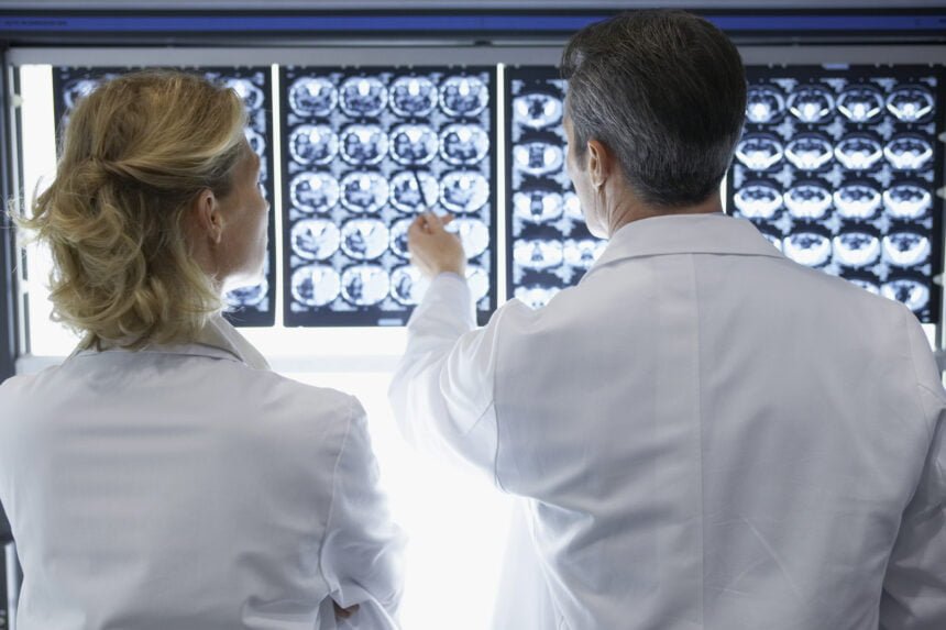
If you pay a visit to the radiology department as part of your treatment process it is highly likely that the medical imaging information created will be in DICOM image format. Digital Imaging and Communications in Medicine (DICOM) is the recognized international standard that allows medical imaging data to be transmitted and saved in a variety of different formats.
You can convert DICOM to PDF, for instance, so that radiologists and other medical professionals can liaise with each other and share results with ease.
Things were not always that straightforward before DICOM became the default standard for storing, printing, displaying, and printing medical images. Previously, results were displayed as two-dimensional film scans. These had to be stored inside film jackets, which was very limiting and not very efficient when it came to sharing and delivering medical imaging results with other relevant departments and people.
DICOM has proved to be a game-changer. It is a standard that has proved to be revolutionary and transformational that has fundamentally altered the radiology industry. It can play a huge role in fighting cancer.
Here is a look at what DICOM is all about and how it can be used to improve functionality and efficiency in the world of radiology.
DICOM image format explained
Digital Imaging and Communications in Medicine (DICOM) is fully recognized as the standard bearer and go-to solution for radiologists when they want to transmit, store, retrieve, print, or process medical imaging information.
One of the big reasons why it is considered such a useful and versatile imaging resource is the fact that you can source DICOM image files from a plethora of different modalities.
As well as being able to save the file as a DICOM file with a standard “.dcm” extension it is also possible to save the files in a variety of different formats such as JPEG and PDF, for example.
As you might imagine, being able to share medical images in numerous different formats makes it much easier to provide these images to other departments and medical professionals in a file format that suits their requirements.
It is this adaptability that has helped DICOM to become the recognized communication standard with radiology departments across the globe.
It should be noted that DICOM files are not able to be opened on a personal computer without the user having the relevant DICOM viewer software loaded.There are several viewer software options to choose from. Popular options are Enterprise Viewer and InteleViewer. Although proprietary software can be used to view images this can be a disadvantage as the image can only be viewed in the same location as the hardware.
Patient security is also a major consideration when it comes to storing and viewing medical images. A typical DICOM file will contain a header and image data combined into one file. The file tag usually contains patient information for ease of identification, however, this can be removed from the header if there is a confidentiality issue.
You can anonymize the data when using formats such as JPEG or PDF if you are sharing images for research purposes.
Advanced functionality options
One of the big reasons why DICOM has become so universally embraced by radiology departments everywhere is that it offers some very useful advanced data enhancement options.
The latest DICOM viewer software available will open up a world of possibilities that could prove useful to radiologists as they are able to enhance and manipulate the data to a certain extent.
You can improve the quality of the image for ease of use, for instance, and you can even take measurements, or increase or decrease the radiodensity and radiolucent areas of an image if you want.
All of these enhancements improve the diagnostic and overall viewing experience.
Another notable advanced functionality option is Multiplanar Reconstruction (MPR). This is a revolutionary new way of generating 3D visualizations of scans from a multitude of different angles. This option provides the opportunity to gain some highly advantageous insights that could even improve medical outcomes.
The ability to combine multiple imaging techniques with the help of DICOM takes accuracy in medical diagnosis to new levels.
Options for sharing and exporting DICOM files
The ability to share and export DICOM image files is an integral aspect of the clinical workflow process.
It is relevant to bear in mind that DICOM files tend to be large in size as a result of the data they contain. When you are storing the results of a CT scan, for instance, that alone will be in excess of 30MB.
It helps practitioners manage these files when they have the ability to use either lossless compression or lossy technology to reduce the size of the file without compromising on quality.
Modern DICOM viewers are able to accommodate these options and then convert the compressed file into another format, such as PDF or JPEG.
Why DICOM is seen as an essential part of radiology
As the above overview confirms, the DICOM image format is widely considered to be an indispensable aspect of radiology practices and workflow options.
Being able to view, manipulate, and share medical images with consummate ease delivers a number of notable benefits. DICOM is a robust security-minded option for capturing and storing image data, delivering an increased level of safety as well as some notable cost savings benefits in the process.
Patient care can be improved as a result of using DICOM technology. It is an option that helps improve productivity and efficiency in radiology departments. As well as being seen as a vital tool for aiding more accurate diagnosis it is able to enhance operational efficiency. That has to be good for delivering better patient care and keeping costs under control.
There is little doubt that DICOM technology has had a huge impact in shaping the way the radiology industry works both now and in the future. The ability to standardize image formats such as X-rays and CT scans and make them available in a single viewing option has proved to be revolutionary.
Now you know why DICOM is so widely embraced by radiologists.

